 by
by X Ray Tech
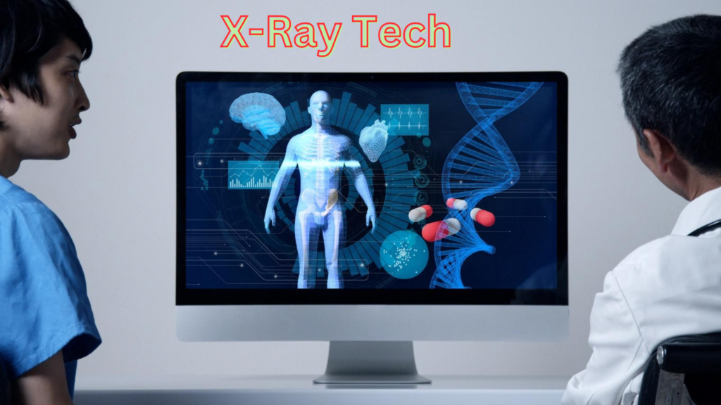
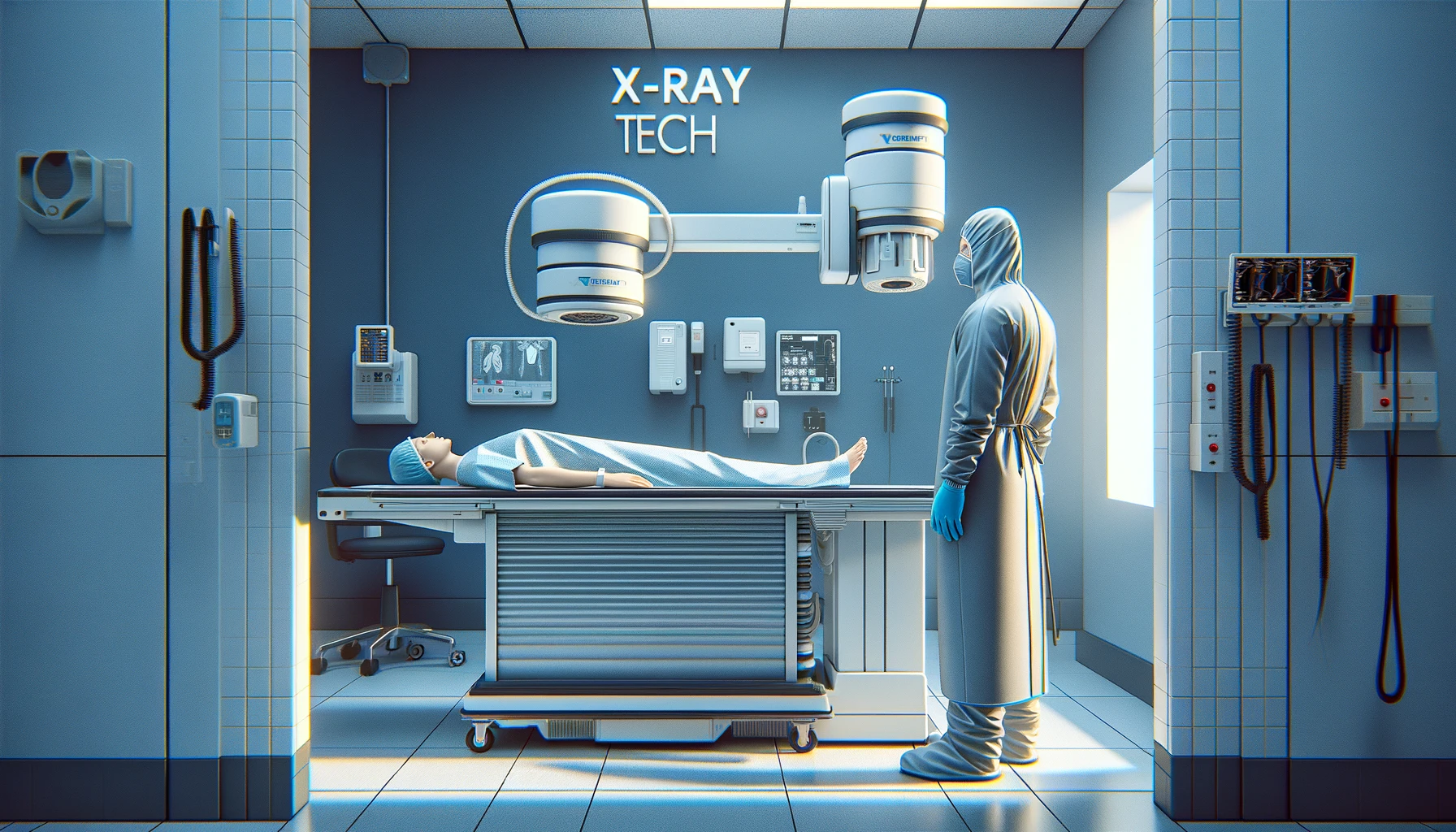
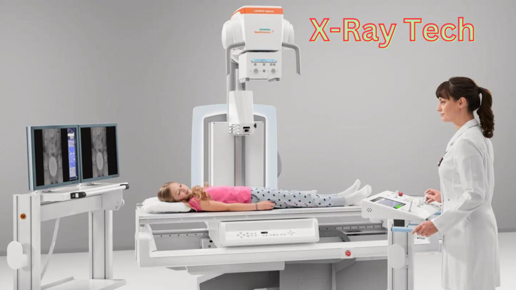
What is the X-Ray Technology
X-Ray technology is fundamental in modern medicine, providing clear images of the body’s interior without invasive procedures. Utilizing electromagnetic radiation, X-rays penetrate various materials, including human tissue, to varying degrees. This variance creates contrasting images on X-ray films or digital sensors, enabling the visualization of bones, specific tissues, and organs. Beyond healthcare, X-ray technology finds applications in security, industrial inspection, and scientific research, demonstrating its versatility and critical role in modern society.
Historical Development and Milestones
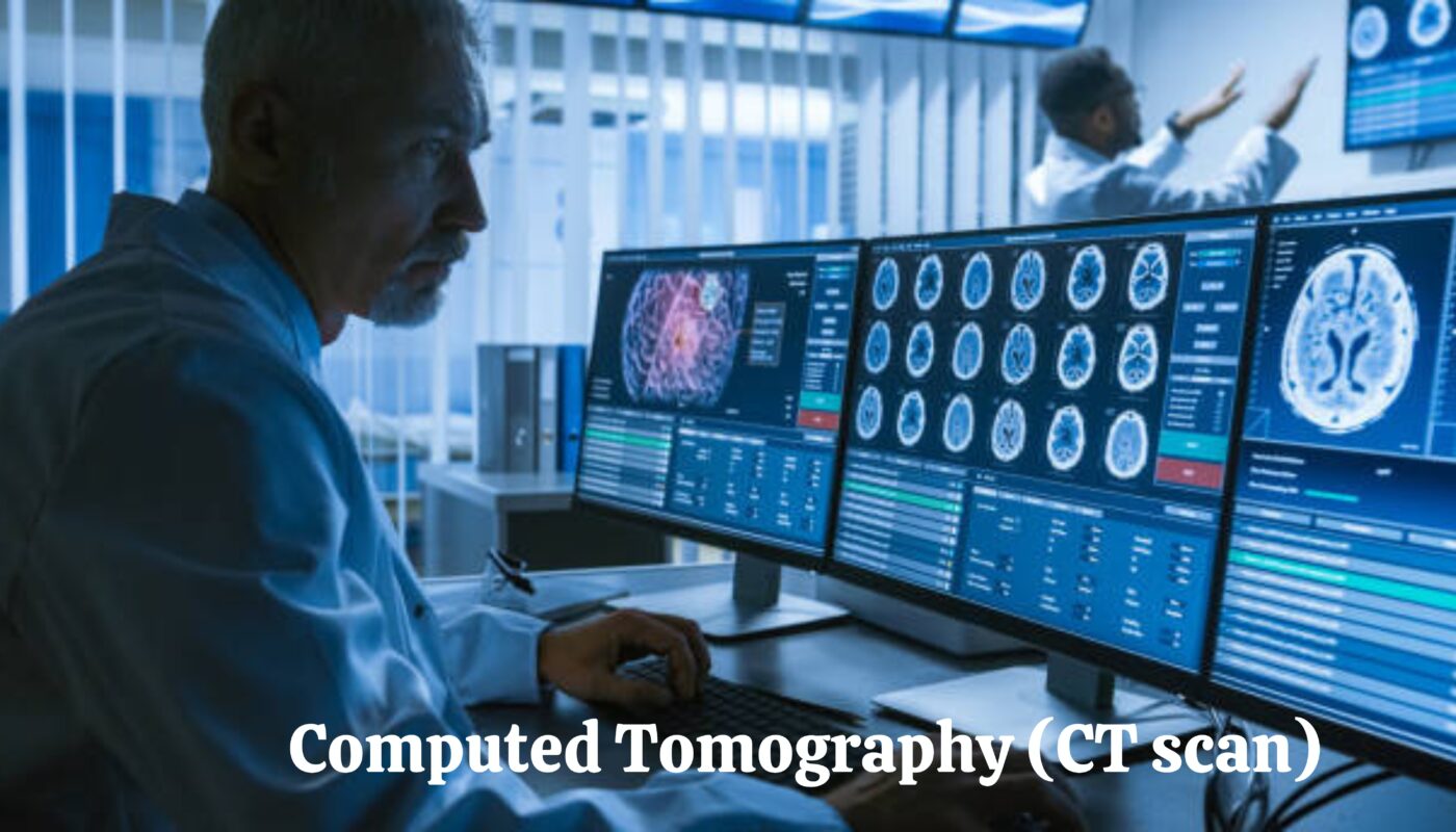
The discovery of X-rays dates back to 1895 by German physicist Wilhelm Conrad Röntgen. He observed that a fluorescent screen in his lab started to glow even though it was not in direct light. This phenomenon was caused by a previously unknown type of radiation, which Röntgen named “X-rays” for their unknown nature. His groundbreaking work earned him the first-ever Nobel Prize in Physics in 1901. Since then, X-ray technology has evolved dramatically. The introduction of computed tomography (CT) in the 1970s, which allows for 3D visualization of the body’s interior, and the development of digital X-ray technology have been significant milestones, vastly improving diagnostic accuracy and patient care.
Importance in Modern Medicine and Industry
In medicine, X-ray technology is indispensable for diagnosing and monitoring numerous conditions, from broken bones to diseases like pneumonia and cancer. It enables timely and accurate diagnoses, significantly impacting treatment outcomes. In industry, X-rays are used for non-destructive testing of materials and structures, ensuring safety and integrity in manufacturing and construction. Furthermore, X-ray technology’s application in security, such as airport baggage screening, underscores its broad utility and importance in maintaining safety and compliance standards.
Basics of X-Ray Technology
How X-Rays Work
X-rays are generated by directing electrons at high speed toward a metal target (typically tungsten). Upon collision, the deceleration of electrons releases energy in the form of X-ray photons. These photons can penetrate different materials, including human tissue. When X-rays pass through the body, they are absorbed by various tissues to varying degrees, creating an image based on the absorption patterns. Denser materials, like bone, absorb more X-rays and appear white on an X-ray film, while less dense materials, such as soft tissues, appear in shades of grey.
Types of X-Ray Imaging Techniques
X-ray imaging techniques have evolved significantly since their discovery, leading to various specialized methods for different diagnostic needs. These techniques leverage the fundamental properties of X-rays to visualize the body’s interior for medical diagnosis, treatment planning, and monitoring. Below are some primary types of X-ray imaging techniques widely used in healthcare.
- Radiography: The most common X-ray imaging used primarily to view bones and specific body structures. Applications include diagnosing fractures and monitoring bone diseases.
- Computed Tomography (CT): A sophisticated technique that combines multiple X-ray images from different angles into a 3D image. It offers detailed views of internal organs, bones, and other tissues, invaluable for diagnosing complex conditions.
- Fluoroscopy: Provides real-time X-ray imaging, ideal for guiding specific medical procedures, such as catheter insertion, and for examining the gastrointestinal tract.
- Dental X-rays: Specifically designed for oral health, these X-rays help dentists detect cavities, monitor tooth roots, and check the jawbone’s health.
Safety Measures and Regulations
Despite the invaluable benefits of X-ray technology, exposure to X-ray radiation poses potential risks, such as increased cancer risk. Therefore, strict safety measures and regulations are in place to minimize exposure. These include using the lowest radiation dose possible for diagnosis, employing lead shields to protect body parts from being imaged, and adhering to regulatory bodies like the FDA and the International Commission on Radiological Protection (ICRP) guidelines. Regular maintenance and calibration of X-ray equipment also ensure the safety and accuracy of diagnostic images.
Advancements in X-Ray Technology
The evolution of X-ray technology has significantly transformed the landscape of medical imaging, providing clinicians and radiologists with tools that offer unprecedented detail and accuracy. The advent of digital imaging, innovations in X-ray equipment, and the integration of artificial intelligence (AI), has enhanced diagnostic precision and streamlined workflows in healthcare settings.
Digital Imaging and Its Benefits
Digital imaging technology marks a pivotal shift from traditional film-based X-rays to digital sensors. This transition offers a multitude of benefits:
- Enhanced Image Quality: Digital images can be adjusted for contrast and brightness, allowing for better visualization of details. This leads to more accurate diagnoses.
- Reduced Radiation Exposure: Digital X-ray systems require less radiation to produce an image of similar contrast to film radiography.
- Efficiency: Digital images are instantly available, eliminating the need for film development and significantly reducing the time from imaging to diagnosis.
- Storage and Sharing: Digital files can be easily stored in electronic health records (EHRs), facilitating secure sharing among healthcare providers and improving collaborative care.
Recent Innovations in X-Ray Equipment
Innovations in X-ray equipment focus on enhancing image quality while minimizing radiation exposure. Some notable advancements include:
- Dual-energy X-ray Absorption Spectroscopy (DEXA): Used primarily to measure bone mineral density for osteoporosis diagnosis, DEXA provides more detailed images with lower radiation doses.
- Digital Tomosynthesis: This technique offers a 3D view of the imaged area and improves the detection of abnormalities by reducing the overlap of structures in the body.
- Portable X-ray Machines: With advancements in portability, X-ray machines can now be used in various settings outside the traditional radiology department, such as inpatient rooms or in the field for sports injuries.
The Role of Artificial Intelligence and Machine Learning
AI and machine learning (ML) technologies are revolutionizing the field of radiology by:
- Automating Routine Tasks: AI algorithms can handle tasks like image sorting and preliminary analysis, allowing radiologists to focus on more complex diagnostics.
- Enhancing Diagnostic Accuracy: Machine learning models trained on vast datasets of X-ray images can help identify subtle patterns not easily visible to the human eye.
- Predictive Analysis: AI can help predict disease risk by analyzing trends and anomalies in a patient’s X-ray images over time.
Google’s Contributions to X-Ray Technology
Google has been at the forefront of integrating AI into medical imaging, demonstrating the potential to enhance diagnostic processes and patient outcomes.
Google AI in Medical Imaging
Google’s AI research in medical imaging focuses on developing algorithms to detect conditions such as lung cancer, diabetic retinopathy, and osteoarthritis from X-ray and other imaging modalities. These AI models are designed to augment the diagnostic capabilities of medical professionals.
Cloud Computing for Image Storage and Analysis
The Google Cloud Platform offers robust solutions for storing and analyzing medical images at scale. Features like:
- Google Cloud Storage: Provides a secure and scalable environment for storing large volumes of imaging data.
- BigQuery: Allows for the fast analysis of large datasets, facilitating research and the development of new diagnostic tools.
Machine Learning Models for Diagnostic Accuracy
Google’s TensorFlow, an open-source ML framework, empowers researchers and developers to build sophisticated models that can improve the accuracy of diagnoses from X-ray images.
Integrating Google Technologies with Xray Tech
Integrating Google technologies into X-ray tech has streamlined imaging processes from acquisition to diagnosis.
Google Cloud Healthcare API for X-Ray Imaging
The Google Cloud Healthcare API enables the seamless integration of imaging data with applications and services, enhancing interoperability and accessibility. This API supports DICOM (Digital Imaging and Communications in Medicine), a standard for storing and transmitting medical images.
TensorFlow for Developing AI Models in Radiology
TensorFlow provides tools for developing and deploying machine learning models that can assist in analyzing X-ray images for diagnostic purposes. It offers a flexible ecosystem for experimenting with novel algorithms and improving existing models.
Collaborations and Research Initiatives
Google actively collaborates with medical institutions and research organizations to explore new applications of its technologies in healthcare. These partnerships aim to accelerate the adoption of AI in medical imaging and to develop solutions that address real-world healthcare challenges.
Case Studies and Real-world Applications
Success Stories of Google Tech in Radiology
Google’s foray into radiology has yielded impressive advancements and success stories, mainly through integrating artificial intelligence (AI) and machine learning (ML) models. One notable example is developing an AI system capable of detecting diabetic retinopathy and macular edema from retinal photographs with accuracy comparable to human experts. This innovation has been pivotal for early detection and treatment, addressing one of the leading causes of blindness globally.
Impact on Healthcare Outcomes
The integration of Google’s technologies in radiology has significantly impacted healthcare outcomes by enhancing diagnostic accuracy, reducing the time for interpretation, and enabling the early detection of diseases. AI-powered tools have also been instrumental in reducing radiologists’ workloads, allowing them to focus on more complex cases and patient care. Moreover, these technologies facilitate better patient management by integrating diagnostic information with electronic health records, leading to more personalized and timely treatment plans.
Future Prospects and Ongoing Research
The prospects of Google tech in radiology are vast, with ongoing research focusing on refining AI models for even more accurate diagnostics across a broader range of conditions. One area of research involves using federated learning, a method that allows AI models to learn from diverse datasets across multiple locations without compromising patient privacy. This approach could significantly enhance the model’s accuracy and applicability in various global health settings. Also, please visit my other post, Digital Therapeutics Alliance.
FAQ on X-Ray Tech
Q1: What is X-ray technology?
X-ray technology is a diagnostic medical imaging technique that uses X-rays to create images of the inside of the body. It helps doctors diagnose, monitor, and treat various medical conditions by providing detailed images of bones, teeth, and soft tissues.
Q2: How do X-rays work?
X-rays work by passing a small amount of ionizing radiation through the body. Different tissues absorb this radiation differently, creating a contrast on the X-ray film or digital sensor. Bones, for example, absorb more radiation and appear white, while soft tissues absorb less and appear in shades of grey.
Q3: What are the common uses of X-ray technology?
Typical uses include diagnosing fractures, detecting certain cancers, examining the lungs, checking for dental problems, and guiding surgeons during specific procedures.
Q4: How long does an X-ray take?
X-ray exams take only a few minutes. More complex studies, like those involving contrast media, may take longer.
Conclusion
Google’s technological innovations have profoundly impacted the radiology field, enhancing diagnostic accuracy, improving healthcare outcomes, and paving the way for the future of medical imaging. The success stories and real-world applications of Google’s AI and cloud computing solutions in radiology underscore the potential of these technologies to transform healthcare. As research continues and these technologies become more integrated into clinical practice, we can anticipate further advancements that will improve patient care and outcomes.
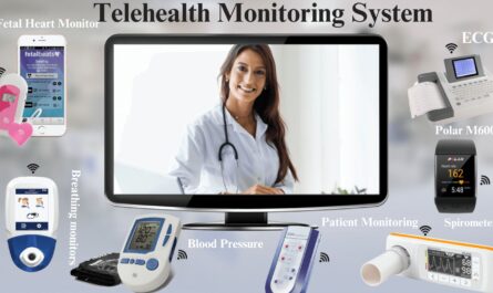
Also, please visit my other post, Telehealth Monitoring System.