27. July 2024
Rigid bronchoscopy

Rigid Bronchoscopy Clearing the Path to Better Breathing

Defining Rigid Bronchoscopy

Rigid bronchoscopy is a medical procedure that provides direct access to the airways and lungs through a rigid bronchoscope—a metal tube inserted through the mouth or nose. Unlike its flexible counterpart, the rigid bronchoscope offers a wider lumen for better airway control, allowing for the performance of various therapeutic interventions such as the removal of foreign bodies, tumor debulking, and stent placement. This procedure is typically performed under general anesthesia in an operating room setting, emphasizing its role in more complex airway management cases.

Historical Background and Evolution of the Procedure

The history of rigid bronchoscopy dates back to the late 19th century, with Gustav Killian performing the first reported bronchoscopy in 1897 to remove a pork bone from a patient’s airway. Since then, the technique has undergone significant evolution, with advancements in technology and anesthesia greatly expanding its applications and safety. The development of the flexible bronchoscope in the 1960s by Shigeto Ikeda introduced a less invasive option for diagnostic purposes, but the rigid bronchoscope has retained its importance for therapeutic interventions where precision and stability are paramount.

The Significance of Rigid Bronchoscopy in Modern Medicine

In modern medicine, rigid bronchoscopy remains an invaluable tool in the diagnosis and treatment of various respiratory diseases. Its ability to provide a secure airway, perform large biopsies, and facilitate the removal of large or complex foreign bodies makes it indispensable in certain clinical scenarios. Moreover, the procedure plays a crucial role in managing emergencies involving airway obstruction, offering life-saving interventions that are not feasible with flexible bronchoscopy. The continued advancements in surgical techniques and instrumentations, such as laser therapy and cryotherapy, further underscore the procedure’s significance in offering minimally invasive options for complex respiratory conditions.

Indications for Rigid Bronchoscopy

Diagnostic Purposes: When Rigid Bronchoscopy is Recommended

Rigid bronchoscopy is recommended for diagnostic purposes in situations where a wide airway access is necessary to obtain large tissue samples for histopathological examination, particularly in cases of suspected lung cancer or when evaluating central airway abnormalities. It is also used when there is a need for direct visualization of the airways to assess anomalies or injuries, such as tracheal stenosis or tracheomalacia, that might not be adequately evaluated with flexible bronchoscopy.

Therapeutic Uses And Other Conditions

Therapeutically, rigid bronchoscopy is the method of choice for managing acute airway obstructions caused by foreign bodies, tumors, or mucous plugs. It is particularly valuable in pediatric cases of foreign body aspiration, providing a safe and effective means of removal. Additionally, the procedure is utilized for dilating narrowed airways, removing cancerous growths, and applying various treatments directly to the airways, such as laser therapy, photodynamic therapy, and stenting to relieve obstruction or seal fistulas.

Differences Between Rigid and Flexible Bronchoscopy

While both rigid and flexible bronchoscopies are invaluable in respiratory medicine, they serve different purposes based on the clinical scenario. Rigid bronchoscopy offers better airway control and the ability to perform more extensive therapeutic interventions, making it suitable for emergency and complex cases. In contrast, flexible bronchoscopy, due to its minimal invasiveness and ability to navigate smaller airways, is preferred for diagnostic purposes and sampling in more distal airway regions. The choice between the two often depends on the nature of the airway problem, the specific therapeutic goals, and the patient’s overall condition, highlighting the complementary roles they play in comprehensive airway management.

Rigid bronchoscopy
Rigid bronchoscopy

3. Preparing for a Rigid Bronchoscopy

Pre-Procedural Assessments and Patient Selection

Before undergoing a rigid bronchoscopy, patients undergo a comprehensive evaluation to assess their suitability for the procedure and to plan the intervention carefully. This assessment typically includes a detailed medical history, physical examination, and review of previous imaging studies, such as X-rays or CT scans, to understand the anatomy and pathology of the airways. Pulmonary function tests and blood work may also be conducted to evaluate the patient’s respiratory status and overall health. Patient selection is based on the indication for bronchoscopy, the potential risks and benefits, and the patient’s ability to tolerate the procedure under general anesthesia.

Patient Preparation: What to Expect Before the Procedure

Patients are usually instructed to fast for a certain period before the procedure to minimize the risk of aspiration during anesthesia. Pre-operative medications may be administered to help with relaxation and to reduce secretions. The healthcare team will also discuss what to expect during and after the procedure, including potential risks and the recovery process. Informed consent is obtained, ensuring that the patient or their legal guardian understands the procedure’s purpose, benefits, and risks.

The Role of the Multidisciplinary Team

A multidisciplinary team approach is crucial for the successful outcome of a rigid bronchoscopy. This team typically includes a thoracic surgeon or pulmonologist with expertise in rigid bronchoscopy, an anesthesiologist experienced in airway management, nursing staff, and, when necessary, other specialists such as interventional radiologists or oncologists. The team works together to plan the procedure, manage any complications, and provide comprehensive post-procedural care, ensuring the best possible outcomes for the patient.

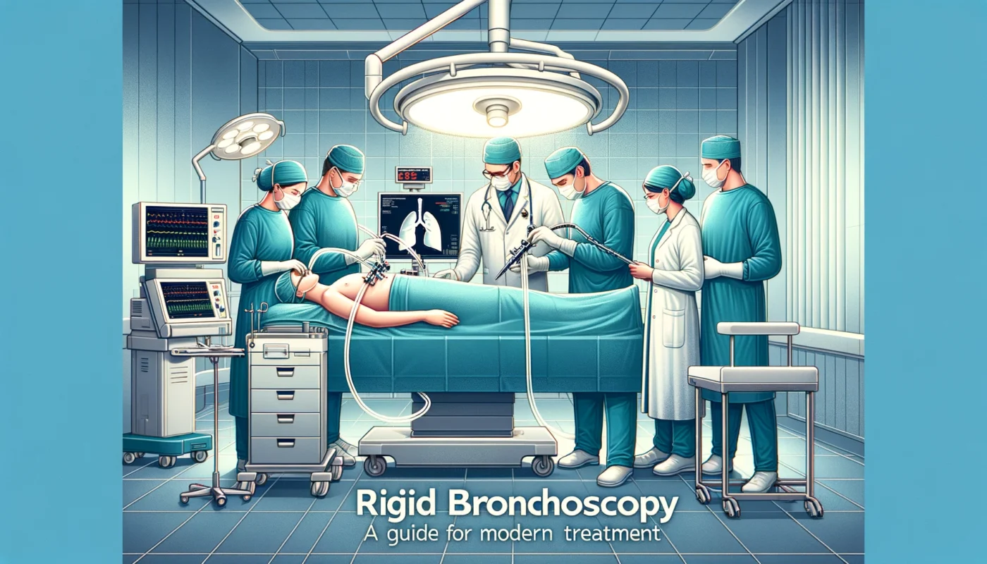
The Rigid Bronchoscopy Procedure

Step-by-Step Guide to the Procedure

  1. Anesthesia and Airway Access: The procedure begins with the administration of general anesthesia and the establishment of a secure airway, often through intubation.
  2. Insertion of the Bronchoscope: Once the patient is sedated, the rigid bronchoscope is carefully inserted through the mouth (or occasionally the nose) and advanced towards the trachea and bronchi.
  3. Inspection and Intervention: The surgeon inspects the airway, identifying any abnormalities. Therapeutic interventions, such as biopsy, foreign body removal, or tumor debulking, are performed as needed.
  4. Completion: After the necessary interventions are completed, the bronchoscope is slowly withdrawn, ensuring the airway remains clear.
  5. Recovery: The patient is closely monitored as they recover from anesthesia and is typically observed for several hours post-procedure for any complications.

Anesthetic Considerations

General anesthesia with muscle relaxation is typically required for rigid bronchoscopy to ensure patient comfort and immobilization, as well as to facilitate control of the airway. Close coordination between the bronchoscopist and the anesthesiologist is essential to manage the airway and ensure adequate ventilation and oxygenation throughout the procedure. Special techniques, such as jet ventilation, may be used to maintain oxygenation while providing the surgeon with enough space to work within the airway.

Tools and Techniques Utilized During Rigid Bronchoscopy

The rigid bronchoscope itself is a straight, hollow metal tube with a light source and a connection for oxygen supply. Various instruments can be passed through the bronchoscope, including forceps for biopsy or foreign body removal, brushes for cytology samples, and lasers or other devices for cutting or cauterizing tissue. Advanced techniques such as cryotherapy, electrocautery, and placement of stents to keep the airway open may also be employed. The choice of tools and techniques depends on the specific goals of the procedure, highlighting the versatility of rigid bronchoscopy in diagnosing and treating a wide range of airway conditions.

Post-Procedure Care and Recovery

Immediate Post-Operative Care

After the completion of a rigid bronchoscopy, patients are moved to a recovery area where they are closely monitored until they fully wake from anesthesia. This includes monitoring vital signs, oxygen saturation levels, and airway patency to ensure the patient is breathing well on their own. Pain management is a crucial aspect of immediate post-operative care, with medications provided as needed to manage any discomfort. The throat may be sore, and the voice hoarse, for which ice chips, throat lozenges, and soft foods are often recommended. Most patients can expect to go home the same day, although some may require overnight observation, especially if the procedure was extensive or if there were complications.

Potential Complications and How They Are Managed

While rigid bronchoscopy is generally safe, it carries potential risks and complications, such as bleeding, infection, airway injury, and reactions to anesthesia. Immediate complications are rare but require prompt recognition and management. For example, significant bleeding can be managed endoscopically using cauterization or topical application of vasoconstrictive agents. Infection risk is minimized with prophylactic antibiotics when indicated. Airway injury, though uncommon, may necessitate additional surgical intervention or prolonged observation. Anesthesia-related issues, such as laryngospasm or aspiration, are managed by the anesthesiology team during and immediately after the procedure.

Long-Term Follow-Up and Rehabilitation

The need for long-term follow-up and rehabilitation varies depending on the reason for the rigid bronchoscopy and any interventions performed during the procedure. Patients undergoing diagnostic bronchoscopy may require follow-up visits to discuss biopsy results and potential further treatment plans. Those who have undergone therapeutic interventions, particularly for conditions like airway stenosis or chronic obstructive pulmonary disease, might need ongoing care to monitor their respiratory status.

It including pulmonary rehabilitation programs to improve lung function and quality of life. Speech therapy may be recommended for patients who have had significant interventions affecting their voice or swallowing. Regular follow-up appointments are crucial to monitor for late complications, evaluate the effectiveness of the treatment, and adjust care plans as necessary. Post-procedure care and recovery after a rigid bronchoscopy are tailored to the individual needs of the patient, taking into account the specific interventions performed and the patient’s overall health. A multidisciplinary approach ensures comprehensive care, addressing both the physical and emotional aspects of recovery to achieve the best possible outcomes for the patient.

Advances in Rigid Bronchoscopy

Technological Innovations and Their Impact

Rigid bronchoscopy has seen significant technological advances that have expanded its capabilities and improved patient outcomes. Innovations such as high-definition video bronchoscopes have enhanced the visual clarity for the operator, allowing for more precise diagnostics and interventions. Laser technology, now a staple in many rigid bronchoscopy procedures, enables accurate cutting and removal of tumors with minimal bleeding, as well as the vaporization of lesions obstructing the airway. Cryotherapy, another advancement, uses extreme cold to effectively treat lesions and manage bleeding.

The development of photodynamic therapy (PDT) offers a minimally invasive option to treat early-stage lung cancer and precancerous lesions in the airways, highlighting the expanding therapeutic scope of bronchoscopy. The integration of navigation technologies borrowed from the fields of radiology and neurosurgery has allowed for more precise localization and treatment of lesions within the lung. These navigational bronchoscopes systems enable the bronchoscopist to reach peripheral lesions that were previously inaccessible with conventional techniques, opening new frontiers in lung cancer diagnosis and treatment. Visit my other post 

Future Directions in Bronchoscope Treatments

The future of rigid bronchoscopy is promising, with ongoing research and development focused on further improving patient care and expanding the range of treatable conditions. One area of intense interest is the use of robotics in bronchoscopy. Robotic-assisted bronchoscopy aims to enhance precision, flexibility, and control during procedures, potentially reducing procedure times and improving safety. Early clinical trials have shown encouraging results, suggesting that robotic systems could soon play a significant role in bronchoscopy treatments. Another exciting development is the exploration of new diagnostic and therapeutic agents delivered directly to the lungs through bronchoscopes.

This includes targeted therapies for lung cancer and advanced gene therapies for genetic lung diseases. The delivery of such treatments directly to the affected areas could maximize therapeutic effects while minimizing systemic side effects, offering a more personalized approach to lung disease treatment. IN the realm of diagnostics, ongoing advancements in imaging and sampling techniques, such as augmented reality (AR) and endobronchial ultrasound (EBUS), promise to further enhance the accuracy of lung disease diagnoses. These technologies aim to provide real-time, detailed images of the lungs and surrounding structures, improving the precision of biopsies and the identification of pathologies.

FAQs on Bronchoscope Treatments

Q1: What is bronchoscopy?
A1: Bronchoscopy is a medical procedure that allows doctors to view the airways and lungs using a bronchoscope, a thin tube with a camera and light.

Q2: Are there different types of bronchoscopy?
A2: Yes, there are two main types: rigid bronchoscopy and flexible bronchoscopy, each used for different purposes and conditions.

Q3: Is bronchoscopy painful?
A3: Patients are usually given local anesthesia or sedation, so they should not feel pain during the procedure.

Q4: How long does a bronchoscopy take?
A4: The procedure typically takes between 30 minutes to an hour, depending on the complexity.

Q5: What are the risks of bronchoscopy?
A5: Though generally safe, risks include bleeding, infection, and reactions to anesthesia, but these are rare.

Conclusion 

Bronchoscopy, particularly the rigid variety, stands as a cornerstone in the diagnosis and treatment of a wide range of pulmonary conditions. With the advent of technological advancements and the potential for future innovations, such as robotic assistance and targeted therapies, bronchoscopy is set to become even more precise, less invasive, and more effective. As medical science continues to evolve, these developments promise to enhance patient outcomes, reduce recovery times, and open new avenues for treating complex lung diseases. Thus, the future of bronchoscope treatments holds great promise for revolutionizing the approach to respiratory healthcare, making it an exciting time for both practitioners in the field and patients benefiting from these advancements.

Leave a Reply

Your email address will not be published. Required fields are marked *