Sonography Technology
Overview of Sonography
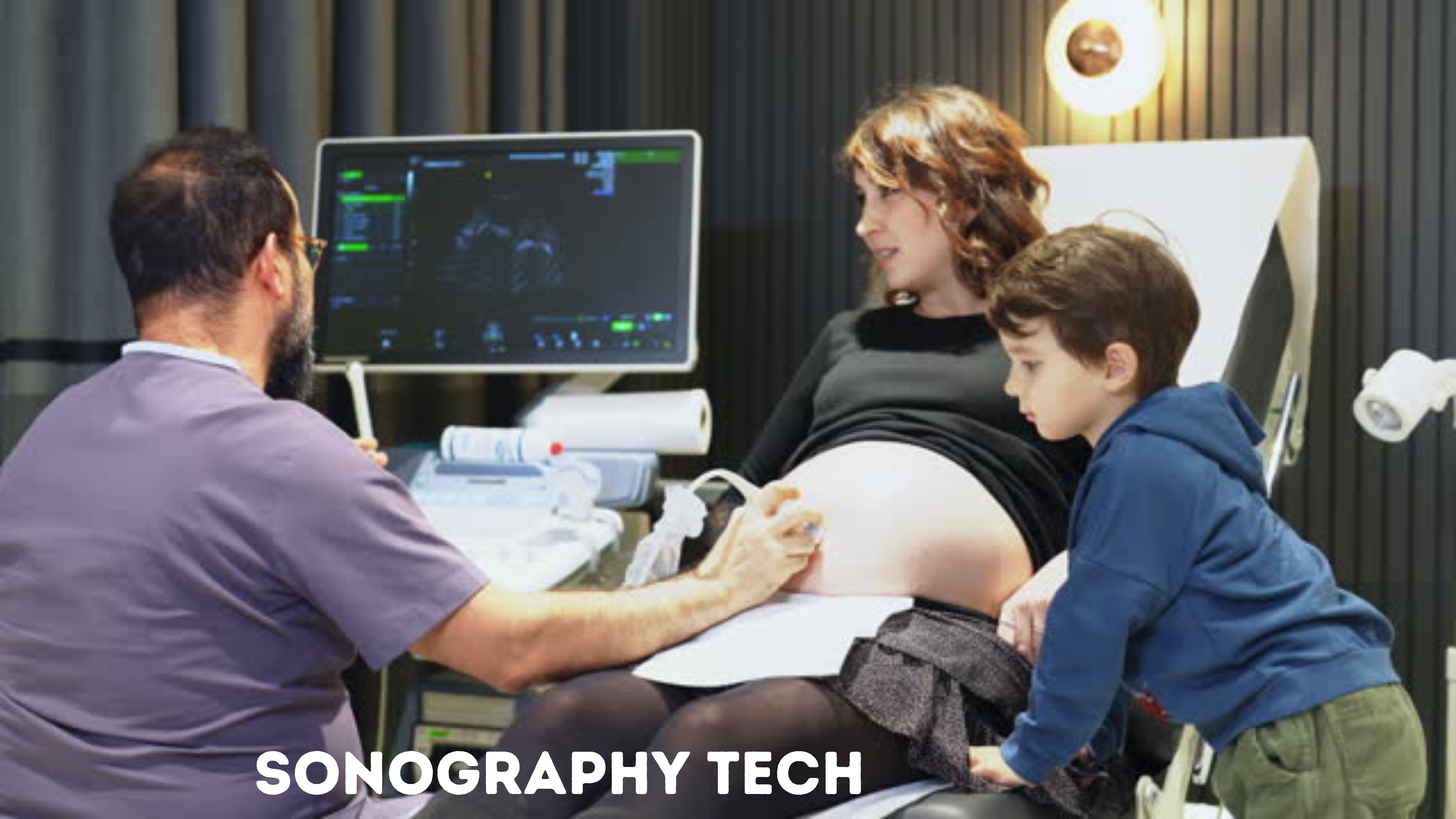
Sonography Tech, often called ultrasound imaging, is a non-invasive diagnostic medical procedure that uses high-frequency sound waves to produce visual images of organs, tissues, or blood flow inside the body. Unlike other imaging techniques, sonography does not use ionizing radiation, making it a safer option for patients, including pregnant women. The images obtained through sonography are crucial for diagnosing and monitoring various medical conditions, guiding surgeries, and assessing fetal development.
Importance in Modern Healthcare
Sonography is a cornerstone for diagnostic precision and patient care in modern healthcare. Its versatility and safety profile have made it indispensable in numerous medical specialties, including obstetrics, cardiology, radiology, and emergency medicine. Sonography helps in the early detection and treatment of diseases, minimizing the need for invasive procedures. Its real-time imaging capability allows healthcare providers to make quick and informed decisions, significantly improving patient outcomes.
Understanding Sonography

How Sonography Works
Sonography operates on the principle of using high-frequency sound waves that echo off bodily structures. A transducer emits these sound waves, which bounce back after hitting tissues, organs, or fluids inside the body. The transducer captures these echoed sounds, and a computer translates them into visual images. The density of the tissues encountered by the sound waves determines the shades of grey in the ultrasound images, with denser structures appearing brighter.
Types of Sonography and Their Uses
As we consider the future of sonography, it’s essential to acknowledge the variety of sonography types and their uses, which are likely to expand and evolve:
- Diagnostic Medical Sonography: Diagnostic Medical Sonography is the most common imaging of the abdomen, reproductive organs, and other internal structures. It is instrumental in diagnosing conditions in the liver, kidneys, uterus, and other organs.
- Cardiovascular Sonography: Focused on the heart and blood vessels, this type helps diagnose heart conditions, assess blood flow, and detect blockages or defects within the cardiovascular system.
- Musculoskeletal Sonography: It is used to visualize muscles, ligaments, tendons, and joints. It assists in diagnosing sprains, tears, and other soft tissue conditions.
The Role of a Sonographer
Sonographers are highly skilled professionals specializing in operating sonography equipment to perform scans. They must deeply understand human anatomy and pathology to capture and interpret images accurately. Sonographers also play a crucial role in patient care, explaining procedures, positioning patients, and ensuring the quality of diagnostic images.
Advancements in Sonography Technology
Recent Technological Innovations
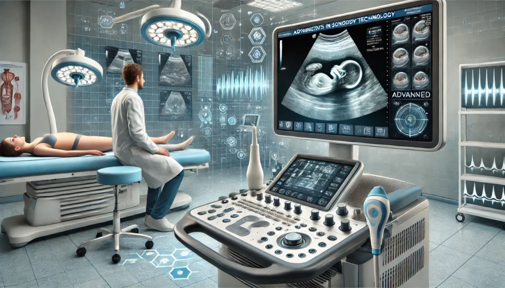
Sonography has seen remarkable advancements, with continuous innovations enhancing image quality and expanding diagnostic capabilities. These include improvements in transducer technology, which allows for higher resolution images, and the development of portable ultrasound machines, making sonography accessible in various settings beyond the traditional clinic or hospital.
3D and 4D Ultrasound Developments
3D ultrasound technology generates three-dimensional images, offering detailed anatomy views that are impossible with standard 2D ultrasound. 4D ultrasounds, an extension of 3D, provide real-time video images showing movement within the body, such as fetal movements or the heart’s beating. These advancements have significantly improved diagnostic accuracy and have become invaluable tools in obstetrics and gynecology, among other fields.
The Impact of AI and Machine Learning
Artificial intelligence (AI) and machine learning are at the forefront of transforming sonography. AI algorithms can analyze ultrasound images much faster and often more accurately than humans, identifying patterns and anomalies that might be missed. This technology also holds the potential to automate parts of the sonography process, reducing the workload on sonographers and improving efficiency in diagnostic procedures. Integrating AI into sonography is paving the way for advancements in personalized medicine, where diagnostic and treatment plans are increasingly tailored to the individual patient’s needs.
Advancements in Sonography Technology

Recent Technological Innovations
The field of sonography has seen significant advancements in recent years, driven by both the demand for more accurate diagnostic tools and the rapid progression of technology. Innovations such as enhanced image clarity, equipment portability, and software integration to analyze images in real time have greatly improved sonography’s capabilities and applications. High-definition imaging allows for more precise, detailed views of soft tissues, blood flow, and small structures, enabling early detection and diagnosis of previously tricky conditions.
3D and 4D Ultrasound Developments
The evolution from traditional 2D ultrasound to 3D and 4D ultrasounds has been a game-changer in medical imaging. 3D ultrasounds create three-dimensional images, providing a static view of the subject, which is particularly useful in obstetrics for examining fetal anatomy for abnormalities. 4D ultrasounds take this a step further by adding the dimension of time, offering moving images that show the fetus in motion. This advancement not only aids in medical assessments but also enhances the bonding experience between parents and their unborn child.
The Impact of AI and Machine Learning
Artificial Intelligence (AI) and Machine Learning (ML) are at the forefront of transforming sonography. These technologies are being used to automate the analysis of ultrasound images, reducing the dependency on human interpretation, which can vary due to experience and fatigue. AI algorithms can assist in detecting patterns not easily visible to the human eye, offering the potential for early detection of diseases such as cancer, liver diseases, and heart conditions. Moreover, AI can streamline workflows by prioritizing cases based on urgency and complexity, ensuring critical cases are reviewed promptly.
Sonography in Practice

Preparing for a Sonogram: What to Expect
Preparation for a sonogram varies depending on the type of examination. For some scans, patients may be advised to drink water and not urinate to fill the bladder, which provides a clearer image of the pelvic organs. For other types, patients might need to fast for several hours beforehand. Generally, patients are instructed to wear comfortable, loose-fitting clothing and may be asked to change into a hospital gown for the procedure. The sonographer will explain the process, how to prepare, and what specific instructions to follow before the appointment.
The Procedure: Steps and Safety Measures
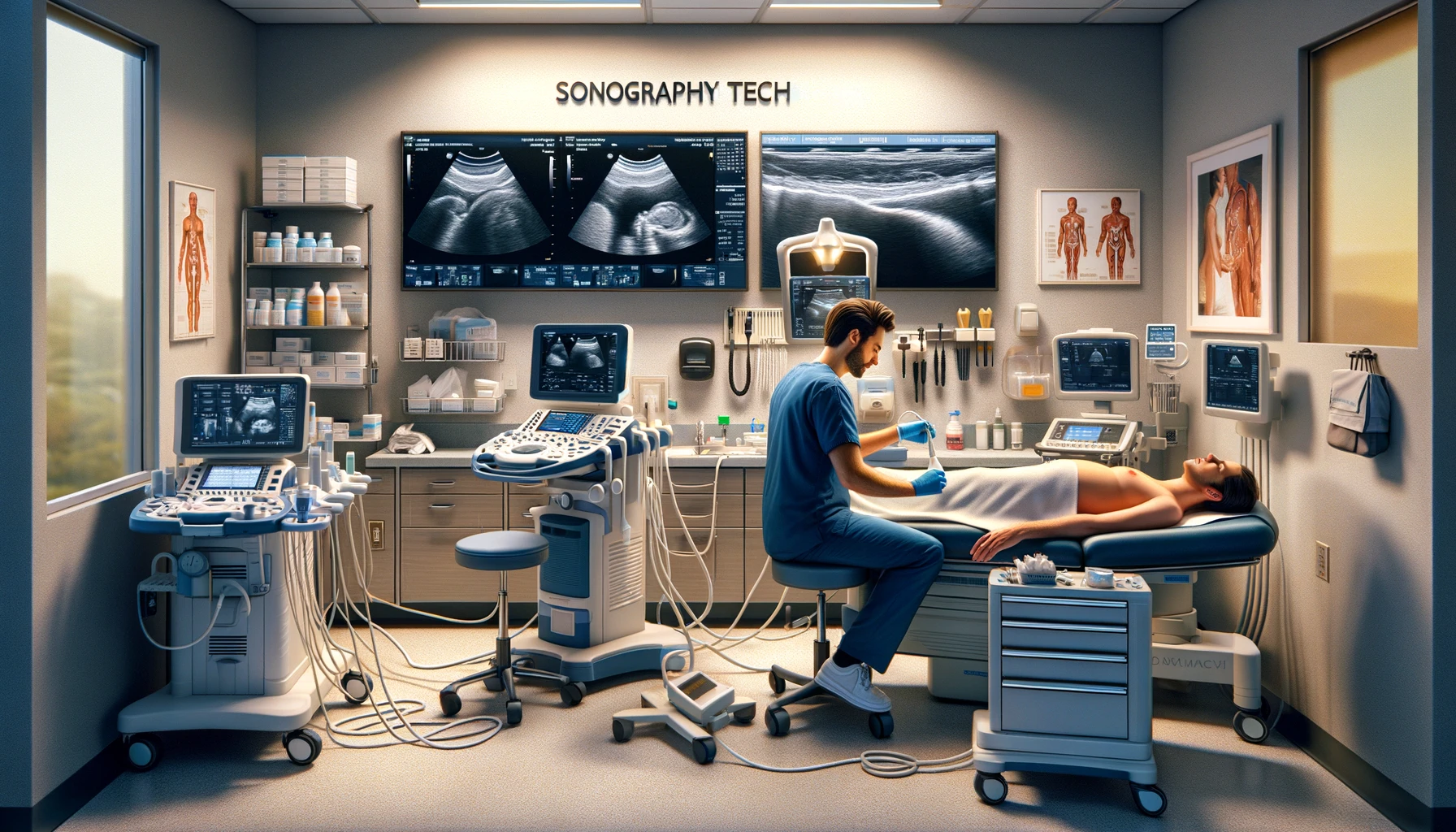
The patient lies on an examination table during the procedure, and a clear gel is applied to the skin. This gel helps transmit the sound waves from the ultrasound probe to the body. The sonographer moves the probe over the skin to capture images of the area of interest. The process is painless and non-invasive, with no ionizing radiation exposure, making it one of the safest diagnostic imaging techniques. The duration of a sonogram can vary, typically between 30 to 60 minutes, depending on the complexity of the examination.
After the Sonogram: Understanding Your Results
Post-procedure, the sonographer may provide immediate feedback. Still, a detailed analysis is usually performed by a radiologist or a specialist who interprets the images and sends a report to the referring physician. The patient will then discuss the results with their doctor, who will explain what has been found and advise on the next steps, including further tests, treatments, or follow-up scans. Understanding the results of a sonogram can be crucial for early intervention and effective management of potential health issues.
The Future of Sonography Technology
As we look toward the future, sonography technology stands on the brink of transformative changes that promise to revolutionize diagnostic imaging. This evolution is driven by advancements in technology, research in medical sciences, and the ever-growing demand for non-invasive diagnostic methods. In this section, we will explore the emerging trends, research areas, and the potential for new applications and improvements in sonography technology.
Emerging Trends and Research Areas
- High-Definition Imaging: Hardware and software advances enable sonographers to produce higher-resolution images, revealing finer details and subtle changes in soft tissues. This high-definition imaging facilitates the early detection of conditions that were previously challenging to diagnose with standard ultrasound technology.
- Integration of AI and Machine Learning: Artificial intelligence (AI) and machine learning are integral to Sonography Tech. These technologies can assist in interpreting ultrasound images more accurately and efficiently, potentially reducing the rate of misdiagnosis and improving patient outcomes. Research focuses on developing AI algorithms that automatically detect abnormalities from ultrasound scans, such as tumours or fetal anomalies.
- Portable and Wearable Ultrasound Devices: The miniaturization of ultrasound devices is leading to the development of portable and even wearable technology. This trend could enable continuous real-time monitoring of patients’ conditions outside of traditional clinical settings. Such devices are especially promising for managing chronic conditions or monitoring pregnancies in high-risk populations.
Potential for New Applications and Improvements
The advancements in sonography technology are not only enhancing current applications but also opening the door to new possibilities:
- Early Disease Detection: Improved image quality and AI-driven analysis can lead to the earlier detection of diseases, such as cancer, by identifying tumors at stages when they are more treatable.
- Telemedicine: Portable and wearable ultrasound devices can facilitate the expansion of telemedicine, allowing healthcare providers to remotely monitor patients and perform diagnostic imaging in remote or underserved areas.
- Surgical Planning and Guidance: Enhanced 3D and 4D imaging can improve surgical planning by providing surgeons with more accurate representations of anatomy. Additionally, real-time imaging during procedures can guide interventions, making them safer and more effective.
- Personalized Medicine: The ability to monitor changes in tissue and organ function over time with high precision can lead to more personalized approaches to treatment, adjusting therapies based on the patient’s response.
Frequently Asked Questions (FAQs) on Sonography Technology
1. What is sonography?
Sonography Tech, also known as ultrasound imaging, is a non-invasive diagnostic procedure that uses high-frequency sound waves to produce images of the inside of the body.
2. How does Sonography Tech work?
Sonography Tech sends sound waves into the body using a small transducer device. The sound waves bounce off tissues, organs, and fluids inside the body, and the echoes are converted into images.
3. What are the common types of sonography?
The most common types include diagnostic medical sonography (for viewing internal organs), cardiovascular sonography (for examining the heart and blood vessels), and musculoskeletal sonography (for assessing muscles, ligaments, and joints).
4. Is sonography safe?
Sonography Tech is considered a safe procedure with no known harmful effects. It creates images using sound waves, not radiation.
5. What is the difference between Sonography Tech and ultrasound?
There is no difference. “Sonography” refers to the technique or process, while “ultrasound” can refer to both the technology and the images produced.
1 thought on “Discover Exciting Advances: Sonography Tech Innovations in 2024”